
Highlight Photo
Developing Sensors for Diagnostics and Monitoring of Clincially Relevant Samples
Figure 4. Bioluminescence response
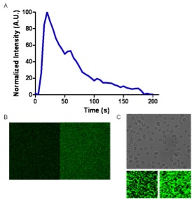
The bioluminescence response observed after the addition of Ca2+ to a solution containing free C5Y82F aequorin with respect to time. B. The image taken of the solution before (left) and after (right) the addition of Ca2+ to the free protein in solution. C. The top imager is the bright field image of the field of view of AML cells bound to the surface of dish with the aequorin conjugated antibody bound to the surface. The bottom two images are the field of view before (left) and after (right) the addition of Ca2+ showing the antibody was bound to the surface of the cells.
Credits: Daniel Scott and Sylvia Daunert