
Highlight Photo
Developing Sensors for Diagnostics and Monitoring of Clincially Relevant Samples
Figure 1. Schematic of simultaneous competitive assay
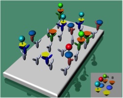
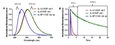
A schematic of the simultaneous competitive assay for IL1?, IL6 and IL8. The grey Y-shaped structures represent the anti-mouse IgG antibodies coated on the microtiter plates. Anti-human IL1?, IL6, and IL8 antibodies are represented by the green, red, and blue Y-shaped structures respectively. Column A in the lower right hand corner represents the interleukin-aequorin fusion proteins while column B represents the free interleukin proteins. IL1? is the orange cone and is attached to the Y82F aequorin mutant paired with ctz f (lime green sphere, detected in the 0-6 s and 515 nm window). The blue ellipsoid represents IL6 and the red sphere Y82F aequorin mutant paired with ctz i (detected in the 6-25 s and 515 nm window). The yellow triangle is IL8 and is fused with the F113W aequorin mutant (cyan sphere, detected in the 0-6 s and 410 nm window).
Credits: Daniel Scott and Sylvia Daunert