
Highlight Photo
Developing Sensors for Diagnostics and Monitoring of Clincially Relevant Samples
Figure 3. Schematic of the devleoped system for detection o AML cells
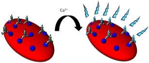
A schematic of the developed system for the detection of AML cells. CD33 (blue) are expressed on the surface of AML cells (red). CD33 is recognized by the anti-CD33 antibody (grey) which is conjugated to C5Y82F aequorin (small spheres on the grey structure). Upon the addition of Ca2+ the aequorin will emit light with the increasing intensity corresponding to an increased number of bound aequorin labeled antibodies.
Credits: Daniel Scott and Sylvia Daunert