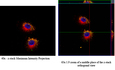

Highlight Photo
Increasing Efficacy for Lung Cancer Chemotherapeutics: Targeted Drug Delivery via the Protocell
Overcoming Macropinocytosis Uptake with a Lipid Bilayer and Increasing Specific Cellular Uptake by Addition of an EGFR Targeting Peptide to the Lipid Bilayer
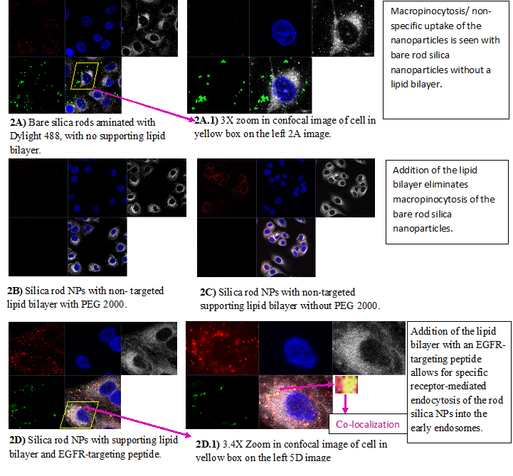
Confocal images of fixed A549 cells incubated with 50 ug of MSNPs in 1 ml of media with 10% FBS. Mid-slice images of the cells for the different groups were taken on a Zeiss 510 META confocal microscope with a 63X oil immersion objective at the University of New Mexicos Fluorescence Microscopy Shared Resource Facility. The stains used for imaging were: a Cell Signaling Technologies EEA1 mAB primary with a secondary Alexa Flour 555 conjugate to stain early endosome antigen proteins (red, top row, 1st column from left); DAPI to stain the cell nuclei (blue, top row, 2nd column from left), Invitrogen Cell Mask deep red 649/666 to stain the cell plasma membranes (light grey, top row, 3rd column from the left), and Thermoscientific Dylight 488 to stain the silica mesoporous rod nano-cores (green, bottom row, 1st column from the left). The merged overlay images of the early endosome, cell nuclei, cell plasma membranes, and the MSNPs are displayed on the bottom row, 2nd column from the left.
Credits: Annikka Jensen – University of New Mexico