

Highlight Photo
Nano-engineered, Ultra Stable, Live Cell Vaccines Against Tuberculosis
Scannin Electron Microscopy and Fluorescent images
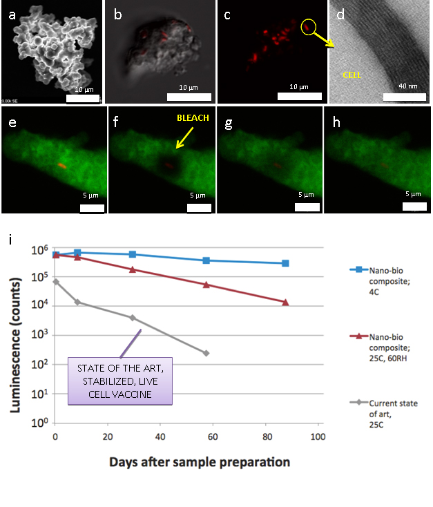
Fig. 1. Individual, dry particles are visualized with scanning electron microscopy (a) and compared to fluorescence images (b,c) of a similar particle, which show the complete encapsulation of E. coli cells expressing a red fluorescent protein. Transmission electron microscopy demonstrates high nanoscopic ordering in the region directly adjacent to an encapsulated cell (d). Fluorescence Recovery After Photobleaching measures the fluid micro-architecture within the dry particle (e-h). An area of known diameter is bleached completely (yellow arrow, f) & the time to recovery of fluorescent molecules is determined. Thermal stability of E. coli encapsulating bio-nano composites was characterized using a viability assay that releases a luminescent molecule in the presence of ATP. This assay was applied to samples aged under refrigerated & accelerated conditions over a three month period (i).
Credits: Patrick Johnson, University of New Mexico
