
Highlight Photo
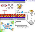
Chemically Directed Immobilization of Nanoparticles onto Gold Substrates
Figure 1. Bright-field and fluorescence images HeLa cancer cells
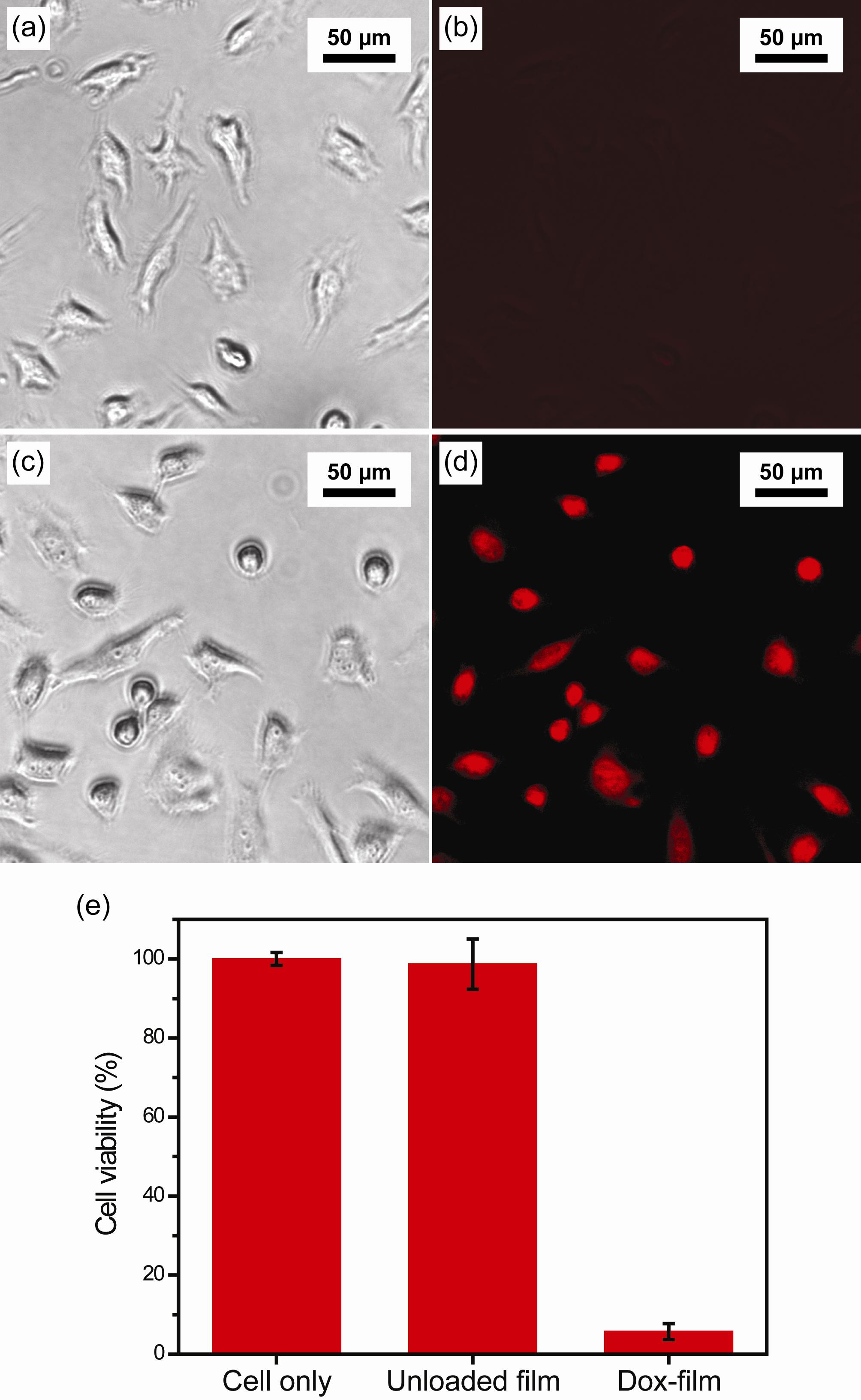
Figure 1. Bright-field and fluorescence images of HeLa cancer cells with unloaded composite films(a, b) and with doxorubicin -loaded dendrimer – nanoparticle films (c, d) after 4 hrs of incubation. (e) In vitro cytotoxicity of unloaded film, and doxorubicin -loaded film using HeLa cancer cells after 26 hrs of incubation.
Credits: Brian Creran, Vincent Rotello