

Highlight Photo
Microbead Array Platforms for High Throughput Screening
Arraying of Microbeads Using Microfluidic Entrapment
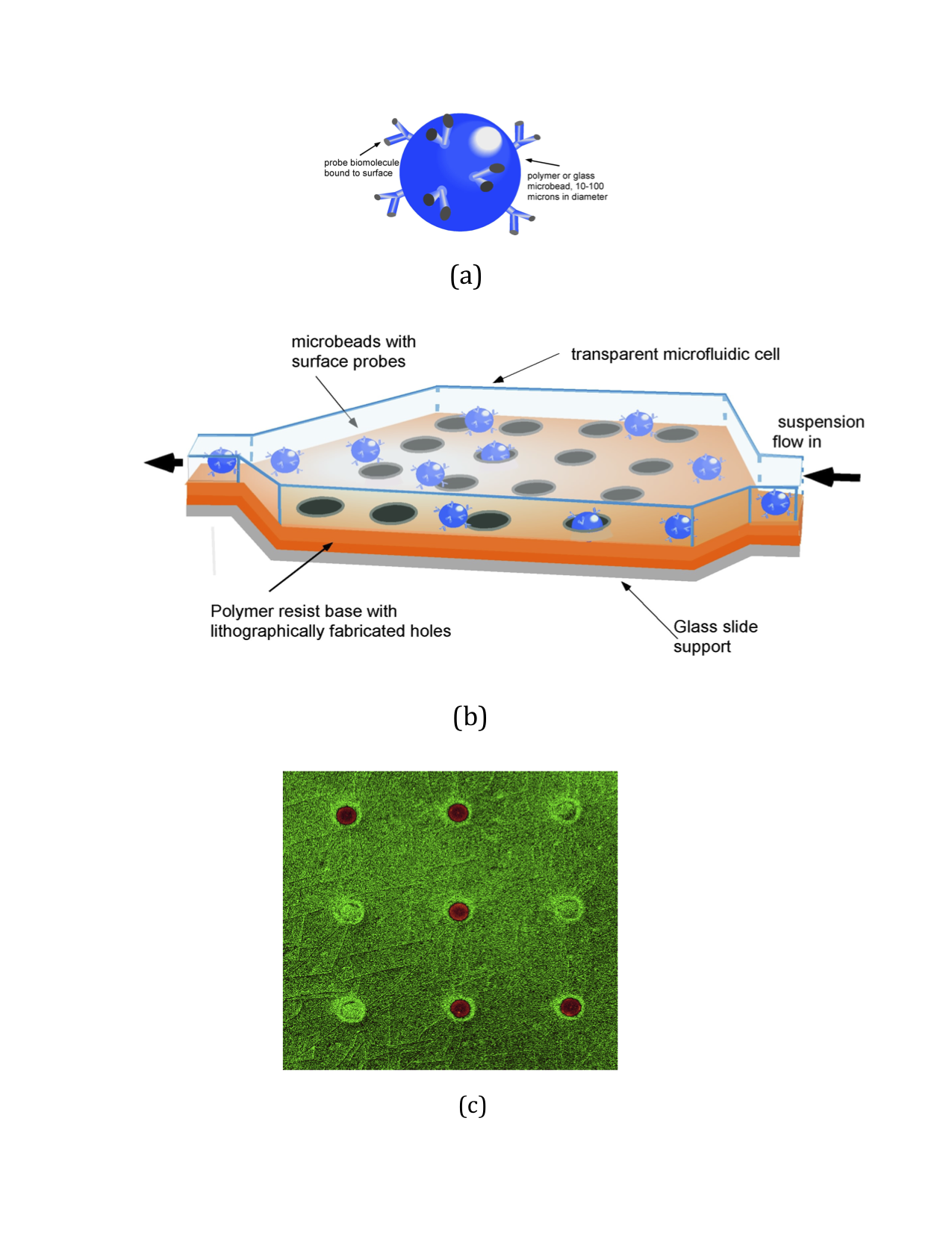
Schematic of (a) the microbead with a probe biomolecule conjugated to its surface, (b) a microfluidic cell obstacle course which uses wells to capture and array the microbead and © fluorescent image of the particles (40 microns in diameter and labeled with a fluorescein dye conjugated to their surfaces) obtained using confocal laser scanning microscopy
Credits: Charles Maldarelli, Levich Institute, City College of New York
