

Highlight
Picket Fence Densities a Determinate of Enhancement vs. Inhibition for Receptor Colocalization
Picket fence density vs. receptor clustering.
Achievement/Results
Michelle Costa, a Trainee supported by the Integrating Nanotechnology with Cell Biology and Neuroscience IGERT program at the University of New Mexico, has shown the effect that receptor confinement has on aggregation and on downstream signaling, shedding light on the duality of previous experimental findings. These results offer insight into an important biological signaling process and provide drug developers with novel targets.
Experimental evidence suggests the cell membrane is compartmentalized into microdomains. In an attempt to understand compartmentalization, a “picket-fence” model was proposed as a potential mechanism; taking into account the receptor-cytoskeletal interactions and their effects on receptor mobility. The effects that receptor confinement has on aggregation as well as downstream signaling remain controversial. Some evidence suggests that receptors confined to a picket-fence leads to aggregation and ultimately an enhancement of signal propagation, while other evidence links receptor confinement to a decrease in dimerization which downregulates signal. Although both scenarios seem to contradict each other, each may hold some truth. Our hypothesis is that the density of these picket fences can enhance receptor aggregation as well as inhibit it.
It has been difficult to determine experimentally the relationship between picket-fence density and receptor aggregation due to poor cell viability. Ms. Costa instead took a computational approach to this problem.
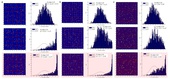
The first question was how did the density of picket fences influence receptor clustering. This was examined comparing the results obtained for three different densities of picket fences, each increasing by a factor of ~2 compared to the previous one. Receptors were randomly placed on the lattice as shown in Fig. 1 (first row) for all distributions. Random distribution was confirmed with the Hopkins test, showing that the data followed a Gaussian distribution, and the Chi-Squared Goodness-of-Fit test, which validated the null hypothesis. Receptors were allowed to diffuse and at a time of 1s (second row) there was a slight right-shift of the data in the Hopkins test. At a time of 2s receptors clustered, as indicated by a dramatic right-shift of the data in the Hopkins test. The chi-squared values no longer supported the null hypothesis, which indicated a non-random, or clustered state.
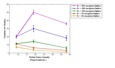
Hypothesizing that receptor concentration may affect the results, simulations were performed for receptor concentrations of 300, 100, 50, and 30 (number of receptors/cell membrane). These results are plotted in Fig. 2. At a low concentration of 30-50 receptors/cell membrane, optimal clustering occurs at the least dense picket fence density of 9 fences/sq micron, where 8~13 clusters are observed. At a concentration between 50 to 100 receptors/cell membrane, a switch within the data occurs, and optimal clustering is observed at the density of 19 fences/sq micron. Simulations at greater receptor concentrations were not preformed, due to computational time, but a switch in optimal clustering is predicted to the most dense 38 fences/sq micron density, at which the cluster size would approximate the number of corrals at this distribution. At a receptor concentration between 200 to 300 (receptors/cell membrane), a limit for the density of 9 fences/micron2 is observed at ~20 clusters, which corresponds to the number of corrals present at this picket fence density.
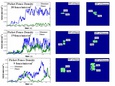
To get a better understanding of how dimerization leads to receptor clustering, a mean square displacement, MSD, of both monomer and dimer for each of the picket fence densities is examined as a function of time and shown in Fig. 3 (left column). Comparing with the theoretical MSD, hindered diffusion is observed for all picket fence distributions. The differences between the monomer and dimer MSD plots are dramatic for the case of the densest distribution of 38 fences/sq micron. The right-hand side in Fig. 3 shows single particle tracking trajectories for both monomer and dimer. Comparing the traces for dimer and monomer, it becomes apparent that dimer is much more confined than its monomer component. These results demonstrate a decrease in receptor mobility when receptors are bound to their partner; supporting oligomer-induced trapping as a mechanism for receptor clustering.
Comparing the three picket fence distributions at a time of 2s (third row in Fig. 1), both the Hopkins as well as the Goodness-of-Fit tests confirm the greatest amount of clustering at the picket fence density of 19 fences/sq micron. The most dense picket fence density of 38 fences/sq micron demonstrated the least amount of clustering, as indicated by both tests; followed by the least dense distribution of 9 fences/sq micron.
At low receptor concentrations (30 to 50 receptors/cell membrane) (Fig. 2), increasing picket fence density has an inhibitory effect on clustering; whereas at normal to high receptor concentrations optimal clustering is observed at a picket fence density of 19 fences/micron2. Increasing from 200 to 300 receptors/cell membrane results in an increase in number of clusters at the picket fence density of 38 fences/micron2. The ability of the cell to synchronize cytoskeletal interactions in conjunction with signaling events has been shown experimentally. Coordinating microdomain densities to regulate cell signaling could prove to be an important mechanism exploited during oncogenesis. The data of Fig .3 show a time delay in clustering, which could activate some signaling pathways while suppressing other pathways. This time delay depends on picket fence density. This concept may be of importance to the activation of ERK, which can lead to either differentiation or proliferation dependent on its transient vs. sustained signal.
Address Goals
One of the hallmarks of oncogenesis is self-sufficiency of growth factors, which leads to an amplification of proliferative pathways. Computer simulation shows that clustering occurs earlier in the case of predimerization, whereas it takes much longer in the case of dimerization. These results support oligomerization-induced trapping as a mechanism for clustering. Such a mechanism is further supported by the MSD plots and the SPT simulations for dimers in comparison to their monomer components.
In summary, these results show how microdomains on the plasma membrane can both inhibit and enhance clustering. When receptor aggregation is enhanced, oncogenic phenotypes such as self-sufficiency of growth factors and amplification of proliferative pathways contributes to the diseased state. Often times, oncogenic events are well coordinated and a mechanism of turning signaling pathways on and off via rearrangement of MSK could facilitate the cancer cell growth.
This work would not have been possible if it weren’t for the collaborative setting of the Integrating Nanotechnology with Cell Biology and Neuroscience (INCBN) IGERT. This fellowship provided the Trainee with the educational and collaborative resources to combine the disciplines of mathematics, computer science, and engineering with the biological sciences, in a very multidisciplinary setting. Such achievements would never have been possible in a single disciplinary setting.