

Highlight
Physics of Membrane Fusion
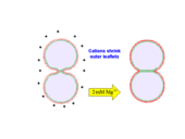
Schematic of divalent cation-mediated vesicle hemifusion.
Achievement/Results
Membrane bilayers composed of amphiphilic phospholipids and proteins are soft materials of great importance to living cells. The fusion of membranes is an essential step in secretion and signaling processes and in intracellular material trafficking. SNAREs (soluble N-ethylmaleimide-sensitive factor attachment protein receptors) and other proteins are thought to mediate the following steps of the fusion pathway: (i) membrane adhesion, (ii) hemifusion, and (iii) complete fusion. Invasion of cells by membrane-enclosed viruses is thought to follow a similar pathway.
Columbia University researchers Prof. Ben O’Shaughnessy and NSF IGERT Traineee Jason M. Warner have mathematically modeled the first two steps on the fusion pathway: adhesion and hemifusion. SNARE-mediated adhesion kinetics and some results for hemifusion kinetics were described in highlights from previous IGERT reporting periods; this highlight focuses on comparing theory with new experimental data.
There is considerable evidence that hemifusion is an intermediate on the pathway to fusion. In the hemifused state the outer leaflets are fused while inner leaflets contact in the hemifusion diaphragm (HD) zone. The hemifused state is metastable: if a hemifusion diaphragm (HD) welding two vesicles grows beyond a critical size, membrane tension tends to expand it. Opposing growth is the interleaflet tension due to compression (expansion) of the outer (inner) membrane leaflets. The model predicts that the equilibrium size is determined by the membrane tension and the vesicle size.
Divalent cations (e.g. calcium, magnesium) are powerful fusion agents used to study hemifusion in model in vitro systems. Cations condense negatively charged membranes, an effect predicted by Warner and O’Shaughnessy to strongly increase the driving force for hemifusion (see Figure 1). The model predicts HD sizes consistent with experimental data (Herrmann group, Humboldt University, Berlin): (i) HD area increases linearly with vesicle size; (ii) HD area increases with vesicle charge.
The model also describes how fast HDs grow. The researchers predict that in the initial phase of HD growth, the HD areal growth rate is constant and determined by initial values of tension and interleaflet tension and the interleaflet drag coefficient (outer monolayers must slide over the inner monolayers to make room for the HD). The models predictions fit well with experimental data (Herrmann group).
These findings are important to understanding the fusion pathway in cells. Understanding the forces which drive or prevent hemifusion elucidates mechanisms for protein-mediated fusion, where SNAREs or other proteins are thought to pull open the hemifusion diaphragm in preparation for complete fusion.
Combination of this theory with experimental results in the future will help understand the mechanisms of biological fusion. In particular, it will be illuminated how the fusion-protein machinery controls HD size on the pathway to fusion. Additionally, this work will shed light on the results from in vivo and in vitro experiments that were previously poorly understood and will motivate future experiments.
Address Goals
This highlight addresses the primary discovery outcome. This research addresses issues key to understanding biological fusion, and additionally, has direct application in biotechnologies (e.g. advanced drug delivery) and medicine (e.g. fighting HIV entry/infection via membrane fusion). Upon addition of these results to the research group’s public website following publication, the contents of this highlight will address the secondary learning outcome by expanding the scientific knowledge of all citizens.
