

Highlight
Investigation of Composite Biodegradable Hydrogel Systems as 3D Porous Scaffolds
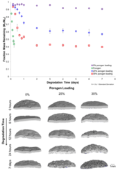
Figure 2: Degradation profiles of the porogen system and composite scaffolds
Achievement/Results
In this project, poly(β-amino ester) (PBAE) biodegradable hydrogel systems were used to create composite hydrogel scaffolds that demonstrated controlled pore opening with degradation. The goal of this research was to create viable scaffolds with controlled pore opening for tissue engineering applications. An outer matrix material was used to provide mechanical support while the porogen microparticles degraded to open a porous network for cell ingrowth (Figure 1). PBAE hydrogel systems have been previously studied and found to have highly tunable properties ideal for biomedical applications (i.e., degradation rate, mechanical compressive moduli). PBAE macromers were synthesized through a step-wise condensation reaction between a diacrylate and amine; the hydrogels were then polymerized using chemically initiated free radical polymerization. Two systems were used, a fast degrading system (~8 hours) was used as the porogen, and a slower degrading system (~3-4 months) was used as the outer matrix. Macromers were synthesized using diethylene glycol diacrylate combined with isobutylamine (outer matrix) or 3-morpholinopropylamine (porogen). Porogen microparticles were created through mechanical grinding and sieved to a range of 250-500 μm for use. These particles were entrapped in the outer matrix during polymerization; systems were referred to by the initial mass loading of porogen. Previous work on these systems has shown that the cytotoxicities examined through MTT analysis are similar to PLGA.
Degradation studies indicated, as expected, an initial phase of rapid degradation due to the loss of the porogen particle mass (Figure 2). Micro-computed tomography (μCT) scans confirmed the creation of a porous network in the scaffold due to the porogen degradation. Porosity analysis indicated greater than 50% porous volume at 7 days in the 35% porogen loaded system. Unconfined compression testing was also completed to observe the mechanical properties of the scaffolds with pore opening. The non-porous scaffold modulus remained the same (>1 MPa) throughout 7 days. The porous scaffolds exhibited a modulus consistent with pore opening and mass loss, the magnitude of the modulus dropped from ~1 MPa to ~0.2 MPa over the course of 7 days due to the opening of the porous network. Cell viability studies were completed on pre-degraded scaffolds to observe if cells were both viable and able to penetrate into the porous network. Scaffolds were degraded for 3 days to remove the porogen particles and open the porous network. A resazurin assay was used to measure viability (Figure 3), and scaffolds were compared to an agarose control because this is hydrogel like in nature. At 24 hours the cells exhibited substantial viability and both the 0% and 35% scaffolds showed values statistically different than the agarose control. This indicates that the cells are viable on the scaffolds and exhibiting viabilities higher than the non-adherent control. Cell tracker was also used to determine if cells were able to move into the pores. Cells were seeded and allowed to incubate for 24 hours, at this point scaffolds were fixed and cryo-sectioned to obtain thin slices for analysis. The fluorescent images were overlaid with the brightfield images to see the cell placement in relation to the pores. It appeared that cells were able to move throughout the scaffold and were concentrated along the pore walls.
Address Goals
This research has advanced the frontiers of knowledge by creating scaffolds from multiple biodegradable hydrogel components that exhibited controlled pore opening. The properties of these scaffolds will benefit tissue engineering as they are ideal for many tissue regeneration applications.
