
Highlight
Discovering how to create appropriate cell-matrix interactions for improving tendon repair
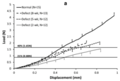
Figure 1a. Average load-displacement failure curve
Achievement/Results
Professor Butler and his team seek to design and fabricate functional tissue engineered “constructs” that surgeons can use to replace load-bearing connective tissues like tendons that connect muscle to bone. Tendons can carry extremely large forces during activities of daily living (ADLs). These forces can approach 40% of failure force during normal ADLs and can rise and fall extremely quickly, often in tenths of a second. It is not surprising that tendon sprains, strains and dislocations represent almost half of all musculoskeletal injuries that occur in the US every year. It is also not surprising that tendon repairs and tendon replacements (grafts from the same individual) never regain the collagen fiber diameter found in the native tissues before injury.
Solving this problem requires a multi-disciplinary strategy from different funding agencies with interdependent and coordinated focus on tissue function, structure, and biology. Past funding from the National Institutes of Health (NIH AR46574- Butler, PI) has been used to determine in vivo tendon forces and the role of adult stem progenitor cells, scaffolds, and mechanical stimulation on tendon repair. Current NIH funding (AR56943- Butler, PI) is being used to determine the biological signals and signaling pathways during normal tendon growth and development. This requires the development of new technologies (e.g. transgenic mice, fluorescent and two-photon imaging, laser capture microdissection and array analysis, and controlled mechanical stimulation during organ culture). This collaborative effort also brings together experts in imaging, molecular biology, materials science, and bioreactors to help solve these problems.
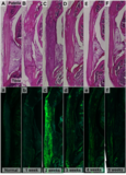
Despite all of these advances, true regeneration of a load-bearing tendon requires a more fundamental understanding of cell-matrix interactions, the precise control of cell phenotype, and spatiotemporal synthesis of appropriate extracellular matrix by these cells that can sustain the large, impulsive forces. The National Science Foundation and the IGERT Program in particular are providing the highly-trained and talented PhD students to work in a team setting to tackle and soon accomplish these objectives. At the University of Cincinnati, Professor Butler and two IGERT trainees (Andrew Breidenbach-current and Nat Dyment-former) as well as Professor Shearn and one former IGERT trainee (Kirsten Kinneberg) are approaching these problems in several unique ways. Kirsten Kinneberg has already demonstrated that using collagen-based sponge scaffolds with mesenchymal progenitor cells found in bone marrow can stiffen a tissue engineered construct (TEC) but do not ultimately improve repair a tendon defect 1. Nat Dyment is also showing that while the cells that fill a tendon defect do upregulate Collagen 1 gene expression (the critical gene for making Type I collagen which is essential for carrying force) by 2 weeks after injury, the resulting tissue that fills the defect is still scar-like with inferior biomechanical properties and histology [Figure 1]2. Both current publications cite NSF IGERT support (0333377).
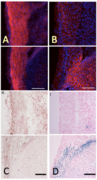
The challenges that we now face are to determine how to direct progenitor cells to synthesize type I collagen, assemble these elements into large diameter fibrils, and discover how to properly insert these into bone through a complex zonal insertion site of collagen, fibrocartilage and bone. To date, no laboratory has been successful in overcoming either of these challenges. Andrew Breidenbach [Figure 2] was recruited to discover how to cause cells to reproducibly form large-diameter fibril in the neighborhood of the cells. Andy is working directly with Nat Dyment and Kirsten Kinneberg to discover what growth factors need to be delivered to mesenchymal progenitor cells in culture, including their timing and interactions. These are based on normal developmental cues for the tendon that show some factors continue to be expressed all along the developing tissue while others are expressed only in the midsubstance or in the insertions [Figure 3] (courtesy of our collaborators, Drs. Christopher Wylie and Chia-Feng Liu). Andrew is examining how to stimulate these progenitor cells with these signals and how these patterns might need to be adapted in the adult healing tendon wound site. Andrew will also be working with Dr. Karl Kadler, a new collaborator at the Welcome Trust at the University of Manchester, UK, who has developed unique 3-D imaging capabilities to rapidly inspect cell shape and collagen fibrillar structure in the neighborhood of the cell. Andrew will contrast two scaffold types (collagen vs. fibrin gels) and cells from three different animal models (mouse, chick, and rabbit) to discover any homology across species. Once achieved, we will optimize growth factors, cell types, and scaffold to significantly improve tissue repair.
References 1. Kinneberg, KRC, Nirmalanandhan, VS, Juncosa-Melvin N, Powell HM, Boyce ST, Shearn JT, Butler DL.
Chondroitin-6-Sulfate Incorporation and Mechanical Stimulation Increase MSC-Collagen Sponge Construct Stiffness. Journal of Orthopaedic Research. 2010; 28(8):1092-1099. (In print) PMC Journal- In Process 2. Dyment NA, Kazemi N, Aschbacher-Smith LE, Barthelery NJ, Kenter K, Gooch C, Shearn JT, Wylie C, Butler DL,
The Relationships between Spatiotemporal Collagen Gene Expression, Histology and Biomechanics Following Full-Length Injury in the Murine Patellar Tendon, Journal of Orthopaedic Research. (In press) PMC Journal- In Process
Address Goals
The achievements described above primarily address the NSF strategic goal of Discovery and the secondary strategic goal of Learning. The research climate that has been established at the University of Cincinnati broadly focuses on expertise in bio-applications of membrane science and technology. The specific highlight described above in the Butler laboratory is concentrating on a biomedical application under this broad biomembrane umbrella. The current NSF trainee (Andrew Breidenbach) and the two former IGERT trainees (Nat Dyment and Kirsten Kinneberg) all seek to discover relevant knowledge in the area of functional tissue engineering. Together they are making important advances related to normal tendon structure and function and the essential features that must be recapitulated when tendon experiences injury. The students are showing the inferior nature of natural healing after injury, including impaired gene expression patterns and subsequent protein deposition and organization, all leading to inferior tendon repair by the introduced and resident host cells.
The students are also part of a very large and diverse group of scientists (bioengineers, molecular biologists, developmental biologists, materials scientists, and surgeons) who are attacking this problem in a very comprehensive fashion. This environment is primarily giving these students the opportunities for new discoveries in genetics, tissue engineering, developmental biology and new materials as well as a learning environment that is unique in the US and around the world. The problem being addressed is complex and multi-faceted and the students can align themselves with experts at the University of Cincinnati and Cincinnati Children’s Hospital (as well as with collaborators at the University of Connecticut [Rowe and Maye], Northeastern University [Ruberti], the Ohio State University [Powell], the University of North Carolina [Banes], and Portland Shriner’s Hospital [Schweitzer]. This broad-based strategy is enriching for these students and should lead to new and exciting fundamental discoveries as well as the likelihood of relevant applied solutions to important biomedical musculoskeletal problems. The interactions among these students (the more senior Nat Dyment and Kirsten Kinneberg mentoring the more junior Andrew Breidenbach) also create a rewarding learning climate, especially given that Nat and Kirsten have already completed the Preparing Future Faculty Program at the University of Cincinnati. This interaction creates a natural learning environment for the “teacher” and the “student.” Taken together, these achievements address both the primary strategic goal of Discovery and the secondary strategic goal of Learning.